- News & information
- About
- History
- George V. Voinovich
- George V. Voinovich Collection
- Calendar
- How to Find Us
- News
- Archives
- Photojournalism Fellowship Project
- Photo Essays
- Current Fellow
- Previous Fellows
- Reports and Publications
- Archives
- Students
- Prospective
- Center for Entrepreneurship
- Environmental Studies
- HTC/Voinovich School Scholars
- Master of Public Administration
- Current
- HTC/Voinovich School Scholars
- Center for Entrepreneurship
- Environmental Studies
- Master of Public Administration
- Alumni
- Contact
- School Leadership
- Strategic Partners Alliance
- Ohio University Public Affairs Advisory Committee
- Ohio University Public Affairs Advisory Committee
- Faculty and Fellows
- Faculty
- Visiting Professors
- Voinovich Fellows
- Professional Staff

Cruciform Nuclear Division
Personal Comments
When I joined the faculty of Environmental & Plant Biology (then it was the Department of Botany) at Ohio University in 1970, Charles E. Miller came to me and described a group of organisms he had studied earlier in his career that had a unique type of nuclear division, "cruciform division" (also see Sorosphaera page). After I read his 1958 paper on Sorosphaera , we set off to Chapel Hill, NC, to collect Veronica with galls for me to prepare for TEM.
The trip was educational for me in several ways. Since much of our trip was through West Virginia before the WVa Turnpike was updated, I got to see a lot of Appalachia for the first time, which was quite an experience for a beginning faculty member who had spent his youth in the rich farmland regions of Wisconsin and Iowa. We made our trip one week after the Buffalo Creek Flood in West Virginia (February 26, 1972), so it was very sobering to see how the tragedy could have occurred as we snaked our way through West Virginia on back roads. Charlie pretty much kept me entertained with stories about his graduate days at the University of North Carolina and early positions in his career, including all the politics at Ohio University at the time. When we arrived in Chapel Hill, Charlie introduced me to his graduate advisor, John Couch; current mycology colleague at UNC, Lindsay S. Olive; and systematist, Al Radford. Lindsay was interested in our project to look at Sorosphaera with the TEM, and eventually incorporated some of our electron micrographs in his book, The Mycetozoans . Al pointed us to several locations on the UNC campus where we could find Veronica infected with Sorosphaera .
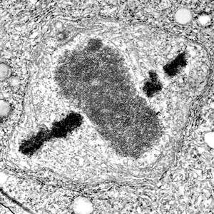
Since our only references were from light microscopy and Keskin's brief paper on Polymyxa , we did not know what to expect for our TEM observations on Sorosphaera . Once I saw centrioles and the other features we eventually described for cruciform divisions in Sorosphaera , I was really pleased that Charlie had introduced me to the group. When I showed him some of the first images of Sorosphaera I took with the TEM (e.g., the image on this page), he decided that he was going to start working on the plasmodiophorids again in addition to his current research at the time on the chytrids.
Images of Cruciform Nuclear Division
- Diagramatic summary of cruciform division
- LMGs comparing cruciform divisions to plant chromosomes
- Tetramyxa cruciform divisions, TEMG
- Cruciform division in Plasmodiophora , TEMG
- Squash of Sorosphaera cruciform divisions, LMG
- Squashes of Sorosphaera cruciform vs. noncruciform divisions, LMG
- Stereo views of Sorosphaera cruciform divisions, CSLM
- Sorosphaera metaphase cruciform divisions, TEMG
- Cruciform nuclear division in Sorosphaera , TEMG
- Cruciform division (anaphase) in Sorosphaera , TEMG
- Sorosphaera cruciform division emphasizing membranes, TEMG
- Sorosphaera cruciform division emphasizing RNP, TEMG
- Cruciform divisions in Hillenburgia sporangial plasmodium on watercress, TEMG
- Cruciform division in Hillenburgia sporangial plasmodium on watercress, TEMG
- Hillenburgia on watercress cruciform division (anaphase), TEMG
References for Cruciform Nuclear Division
- Braselton, J. P., C. E. Miller, & D. G. Pechak. 1975. The ultrastructure of cruciform nuclear division in Sorosphaera veronicae (Plasmodiophoromycete). Amer. J. Bot. 62: 349-358.
- Dylewski, D. P., J. P. Braselton, & C. E. Miller. 1978. Cruciform nuclear division in Sorosphaera veronicae . Amer. J. Bot. 65: 258-267.
- Dylewski, D. P. & C. E. Miller.1983. Cruciform nuclear division in Woronina pythii (Plasmodiophoromycetes). Amer. J. Bot. 70: 1325-1339.
- Garber, R. C. & J. R. Aist. 1979. The ultrastructure of mitosis in Plasmodiophora brassicae (Plasmodiophorales). J. Cell Sci. 40: 89-110.
- Horne, A. S. 1930. Nuclear division in the Plasmodiophorales. Ann. Bot. 44: 199-231.
- Keskin, B. 1971. Beitrag zur Protomitose bei Polymyxa betae Keskin. Arch. Mikrobiol. 77: 344-348.
Contact Information:
(740) 593–9381 | Building 21, The Ridges
Ohio University Contact Information:
Ohio University | Athens OH 45701 | 740.593.1000 ADA Compliance | © 2018 Ohio University . All rights reserved.
